3 December 2008

Meningitis in Children


Meningitis is an infection in the nervous system. It can be caused by bacteria and virus or complication from neurosurgery, trauma, and infection in sinus or ears.

Meningococcal meningitis occurs in epidemic form and transmitted by droplet infection from nasopharyngeal sections. Viral meningitis is associated with viruses such as mumps, paramyxovirus, herpes virus, and enterovirus.

The diagnoses of meningitis is made by testing cerebrospinal fluid that shows increased pressure, cloudy, high of protein, and low glucose.

Signs and Symptoms:
  • There are no classic signs and symptoms. It depends on the type, child age, and duration of illness.
  • Vomiting and diarrhea
  • Fever, chills
  • Nuchal rigidity
  • Poor feeding or anorexia
  • Alter level of consciousness
  • Bulging anterior fontanel in infant
  • Muscle or joint pain
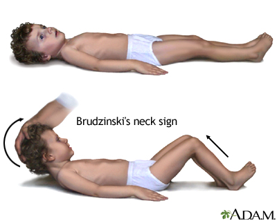
  • Kernig's sign and Brudzinski's sign


Nursing Intervention:
  • Isolation for at least 24 hours after antibiotic initiated
  • Administer antibiotics as prescribed
  • Assess neurological and cardiovascular system
  • Monitor intake and output

Reye's Syndrome


Reye's syndrome is acute encephalopathy follows a viral illness. It is characterized by cerebral edema and fatty changes in the liver. The disease is associated with administration of aspirin. The main goal of treatment is to maintain effective cerebral perfusion and control increasing Intra Cranial Pressure.


Signs and Symptoms:
  • History of viral illness 4-7 days before the onset of symptoms
  • Nausea and vomiting
  • Progressive neurological deterioration
  • Malaise
Nursing Intervention:
  • Assess and monitor neurological status
  • Monitor LOC and signs of increased ICP
  • Provide rest and decrease stimulation
  • Monitor intake and output
  • Monitor liver function
  • Monitor for signs of bleeding and impaired coagulation

Spina Bifida


Spina bifida is a defect in central nervous system. It occurs as a result from neural tube failure to close during embryonic development.

Type of Spina Bifida
1. Spina Bifida Occulta:
Posterior vertebral arches fail to close in the lumbosacral area. Spinal cord remains intact and usually is not visible. Meninges are not exposed on the skin surface and neurological deficit are not usually present.

2. Spina Bifida Cystica
The vertebra and neural tube close incomplete resulting in a saclike protrusion in the lumbar or sacral area. The defect includes meningocele, myelomeningocele, lipomeningocele, and lipomeningomyelocele.

3. Meningocele
The protrusion involves meninges and a saclike cyst that contains CSF in the midline of the back. Spinal cord is not involved and neurological deficits are usually not present.

4. Myelomeningocel
The protrusion involves meninges, CSF, nerve roots, and spinal cord. The sac is covered by a thin membrane that is prone to leakage or rupture. Neurological deficit are evident.


Signs and Symptoms:

  • Visible spinal defect
  • Flaccid paralysis of the legs
  • Hip and joint deformities
  • Altered bladder and bowel function
  • Specific signs and symptoms depend on the spinal cord involvement

Nursing Intervention:
  • Assess the sac and measure the lesion
  • Assess neurological system
  • Assess and monitor for increasing ICP
  • Measure head circumferences
  • Protect the sac, cover with a sterile, moist (normal saline), nonadherent dressing and change the dressing every 2-4 hours
  • Place patient in prone position and head to one side
  • Use antiseptic technique
  • Assess and monitor the sac for redness, clear or purulent drainage, abrasions, irritation, and signs of infection
  • Assess for hip and joint deformities
  • Administer medication: antibiotics, anticholinergics, and laxatives as prescribed