Spina bifida is a defect in central nervous system. It occurs as a result from neural tube failure to close during embryonic development.
Type of Spina Bifida
1. Spina Bifida Occulta:
Posterior vertebral arches fail to close in the lumbosacral area. Spinal cord remains intact and usually is not visible. Meninges are not exposed on the skin surface and neurological deficit are not usually present.
2. Spina Bifida Cystica
The vertebra and neural tube close incomplete resulting in a saclike protrusion in the lumbar or sacral area. The defect includes meningocele, myelomeningocele, lipomeningocele, and lipomeningomyelocele.
3. Meningocele
The protrusion involves meninges and a saclike cyst that contains CSF in the midline of the back. Spinal cord is not involved and neurological deficits are usually not present.
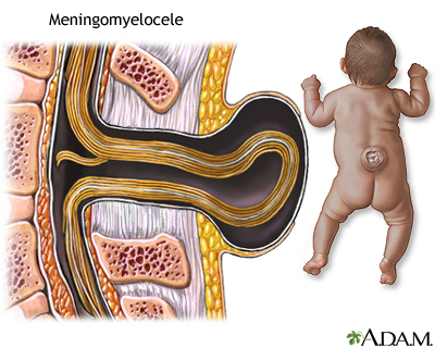
4. Myelomeningocel
The protrusion involves meninges, CSF, nerve roots, and spinal cord. The sac is covered by a thin membrane that is prone to leakage or rupture. Neurological deficit are evident.
Signs and Symptoms:
- Visible spinal defect
- Flaccid paralysis of the legs
- Hip and joint deformities
- Altered bladder and bowel function
- Specific signs and symptoms depend on the spinal cord involvement
Nursing Intervention:
- Assess the sac and measure the lesion
- Assess neurological system
- Assess and monitor for increasing ICP
- Measure head circumferences
- Protect the sac, cover with a sterile, moist (normal saline), nonadherent dressing and change the dressing every 2-4 hours
- Place patient in prone position and head to one side
- Use antiseptic technique
- Assess and monitor the sac for redness, clear or purulent drainage, abrasions, irritation, and signs of infection
- Assess for hip and joint deformities
- Administer medication: antibiotics, anticholinergics, and laxatives as prescribed
No comments:
Post a Comment